About the NONCOMPACT study
In a large cohort of adult patients with suspected NCCM we will perform in-depth phenotyping, including clinical information, pedigree data, genetics, echocardiography and MRI, and follow participants for up to 3 years. We will apply machine-learning based analytics to develop predictive models and compare their performance to currently used models and treatment criteria. Secondly, in a subset of participants we will perform high-resolution cardiac CT for detailed structural characterization of the myocardial wall. We will investigate associations between myocardial structure and regional contractile function, as assessed by echo and MRI.
The aim of this study is to identify a structural signature associated with pathological non-compaction and improve developed risk prediction models. Discovery of pathological structural signatures through innovative imaging techniques, in relation to myocardial contractility, will advance our understanding of NCCM.
Inclusion criteria
- At least 18 years old
- Increased trabeculations of the left ventricle seen by echocardiography
- A cardiac MRI scan was previously performed or is planned for clinical reasons
General exclusion criteria
- Complex congenital heart disease, neuromuscular disorders, increased trabeculations of the right ventricle alone
- Inability to safely undergo cardiac MRI (only applies if an MRI scan has not yet been performed), for example: pacemaker or ICD, contrast allergy, irregular heart rhythm, severe shortness of breath
Exclusion criteria for CT scan (if contra-indications to CT exist participation in the general study is still possible)
- Age below 21 years
- Decompensated heart failure, or otherwise clinically unstable
- BMI>40 kg/m2
- Pregnancy (or cannot be ruled out)
- Known iodine contrast medium allergy
- Kidney dysfunction: eGFR<45 ml/min
- Thyroid disease: toxic multinodular goiter, Graves' disease, Hashimoto's thyroiditis
Supported by
What is non-compaction of the left ventricle?
Left ventricular non-compaction (LVNC), also referred to as non-compaction cardiomyopathy or increased trabeculation of the left ventricle, is a poorly understood disorder characterized by a left heart chamber with a prominent inner layer of loose myocardial tissue. The condition has been associated with heart failure, stroke and arrhythmia. However, for most patients with increased trabeculations there are no negative consequences.
Why do we study LV non-compaction?
LV non-compaction is more and more often diagnosed, partially because of the improved quality of imaging techniques. A small proportion of patients with increased trabeculations develop complications, but in the vast majority the condition does not cause problems in the future. Currently we are not well able to tell which type of non-compaction is harmless and which one requires measures to prevent complications. For a growing group of patients diagnosed with LV non-compaction there is a need for better risk stratification to appropriately allocate (or safely withhold) impactful preventive measures such as ICDs and anticoagulation (blood thinners).
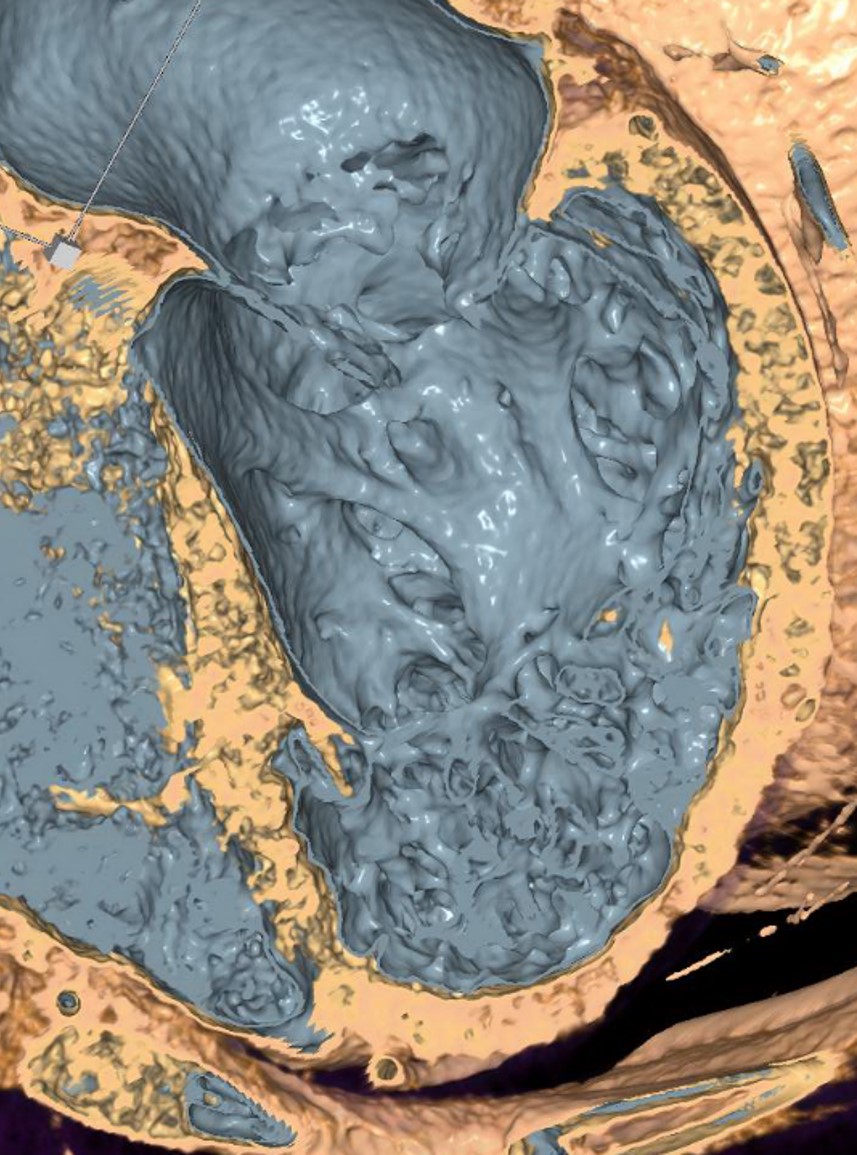
3D structure of the LV wall
3D Rendered inside view of the left ventricle, which shows the complex trabecular structure of the ventricular wall.